

Did you know that roughly 55% of women report an adverse change in breast shape following pregnancy, yet most walk into a consultation without ever hearing the phrase “glandular-to-skin-envelope ratio”? According to Aesthetx, a leading plastic surgery center medically reviewed by Dr. Kamakshi R. Zeidler, post-lactational involution involves the regression of secretory lobules through increased apoptosis, phagocytosis of residual milk, and stromal remodeling — a biological event that transforms your breast from the inside out (Aesthetx, Post-Pregnancy Breast Changes, 2026). This gives rise to the single most consequential decision in your mommy makeover: breast lift + implant vs. lift-only.
That decision cannot be made by flipping through before-and-after photos alone. It demands a glandular volume assessment — a precise reading of how much functional breast parenchyma you still carry versus how much skin envelope your pregnancy left behind. Underestimate your glandular reserve, and you wind up over-augmented. Overestimate it, and you walk away with a lift that looks hollow within six months. This guide delivers the 2026 framework that settles the breast lift + implant vs. lift-only question once and for all, built from Dr. Sibel Atalay’s 29-year clinical practice in Antalya.
The Anatomy of Post-Pregnancy Breast Involution: What Actually Happens Inside
Understanding the breast lift + implant vs. lift-only debate starts with grasping what post-pregnancy breast involution does on a cellular level. During pregnancy, your ductal system expands. Glandular tissue increases in density and volume. Your skin envelope stretches to accommodate this growth. Then you wean. The milk-producing tissue regresses, partially replaced by adipose tissue. The result: reduced volume, skin laxity, and a downward shift in nipple position.
Aesthetx confirms that breastfeeding itself has not been found to be an independent risk factor for breast ptosis. The pregnancy itself drives the aesthetic changes. This distinction matters profoundly. Women who pump and women who never lactate still face the same involution pattern. The glandular tissue retracts regardless of feeding method, leaving behind an envelope that no longer matches its contents.
The Glandular-to-Skin-Envelope Ratio: Your 2026 Decision Metric
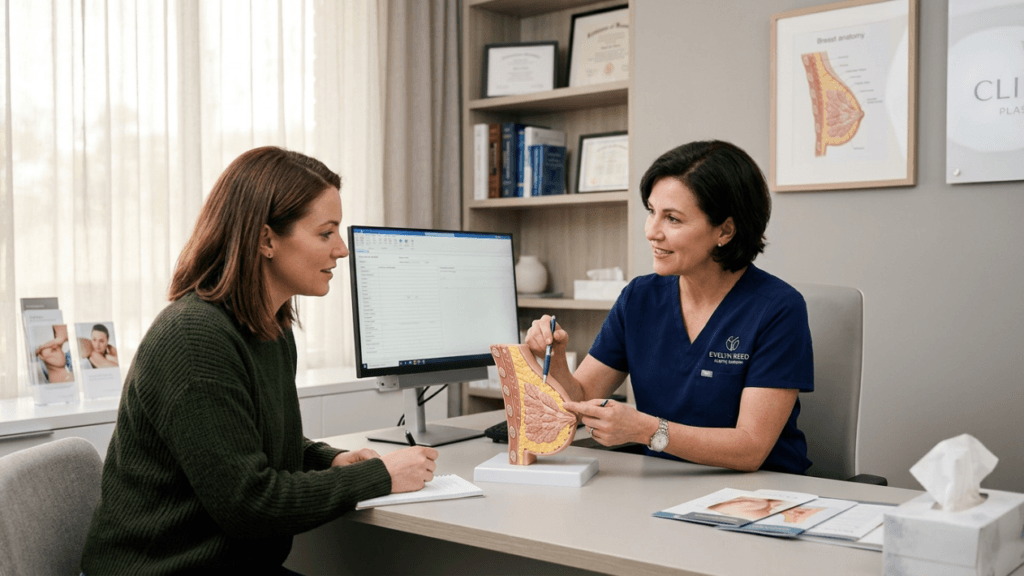
The skin envelope ratio quantifies the mismatch between your remaining glandular volume and the skin that holds it. Think of it as measuring a deflated balloon: the balloon skin still exists, but the air inside has vanished. When you consult with our board-certified surgeons, this ratio becomes the Rosetta Stone for your surgical plan.
Here is the core principle: mastopexy-only vs. augmentation-mastopexy is not a preference question. It is a math problem. If your glandular volume can fill a tightened skin envelope at a size you find acceptable, you need a lift alone. If your glandular reserve falls short — leaving upper-pole hollowing even after skin tightening — an implant becomes necessary.
Calculating the Glandular-to-Skin-Envelope Ratio
Surgeons calculate this ratio through direct clinical examination, assessing three variables:
- Glandular Parenchymal Volume: The amount of functional breast tissue remaining after involution. Measured by palpation of parenchymal thickness at the upper, medial, and lower poles.
- Skin Envelope Surface Area: The total skin surface available, estimated by measuring the base width, base-to-nipple distance, and medial-to-lateral spread of the breast footprint.
- Ratio Output: Divide parenchymal volume by envelope area. Higher ratios suggest sufficient glandular tissue for lift-only candidacy. Lower ratios flag the need for augmentation-mastopexy.
How Ptosis Grade and Implant Need Intersect After Pregnancy
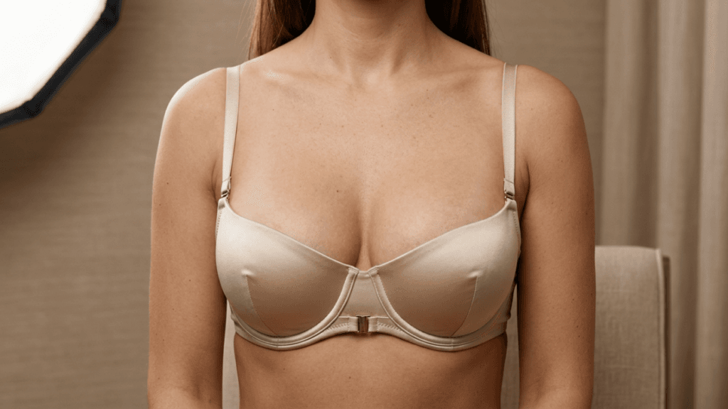
Ptosis severity — measured by the Regnault classification — tells your surgeon where the nipple sits relative to the inframammary fold. But ptosis grade and implant need do not travel in a straight line. A patient with severe ptosis and robust glandular volume may achieve a beautiful result from mastopexy alone. A patient with mild ptosis but severe upper-pole hollowing will almost certainly need an implant.
Consider this real-world pattern seen in Dr. Sibel Atalay’s Antalya clinic: a 34-year-old mother of two presents with Grade II ptosis. Her nipples sit 2 centimeters below the fold. Her glandular tissue is thin at the upper pole but adequate in the lower pole. She requests breast augmentation with implants alone, believing that volume will “lift” the breast. This assumption fails. Implants placed behind a lax skin envelope without tightening the envelope simply push the breast further downward, creating what surgeons call a “double-bubble” deformity. The correct decision is augmentation-mastopexy.
Conversely, a 39-year-old mother of three presents with Grade III ptosis but retains substantial glandular volume in the central and lower poles. Her skin is stretched, yes, but her parenchymal thickness measures robust on examination. A mastopexy alone — reshaping and tightening without any implant — restores her pre-pregnancy contour beautifully. Adding an implant here would overfill the breast, strain the incisions, and accelerate recurrent ptosis.
Mastopexy-Only vs. Augmentation-Mastopexy: The Comparative Framework
The following table breaks down the decision between mastopexy-only vs. augmentation-mastopexy across the variables that genuinely matter to post-pregnancy patients. Study it carefully. This is the decision matrix your consultation should revolve around.
| Assessment Variable | Mastopexy-Only Candidate | Augmentation-Mastopexy Candidate |
|---|---|---|
| Glandular-to-Envelope Ratio | High — sufficient parenchyma fills tightened envelope | Low — parenchyma cannot fill envelope even after tightening |
| Upper-Pole Fullness Pre-Op | Adequate or mild deficit | Significant hollowing, visible concavity |
| Pre-Pregnancy Cup Size | C cup or larger before pregnancy | A or B cup before pregnancy |
| Involution Pattern | Central and lower-pole dominant | Upper-pole dominant (“empty balloon” look) |
| Skin Elasticity | Good to moderate | Poor (multiple pregnancies, significant striae) |
| Patient Volume Goal | Same as pre-pregnancy size | Larger than pre-pregnancy or restores lost fullness |
| Risk of Recurrent Ptosis | Lower (less weight on repaired envelope) | Higher (implant adds weight to tightened skin) |
| Scar Burden | Lollipop or anchor depending on ptosis grade | Anchor pattern virtually always required |
| Revision Rate | 5–8% within 3 years | 8–17% within 3 years (per multiple clinical studies) |
This comparison illuminates a critical blind spot. Most online guides treat the breast lift + implant vs. lift-only question as a matter of desire — “Do you want to be bigger?” — when it should start as a matter of anatomy. The glandular assessment answers the question before personal preference even enters the conversation.
The Post-Pregnancy Involution Patterns That Change Everything
Every mother’s breasts tell a different post-pregnancy story. Recognizing your involution pattern unlocks the correct surgical path. Dr. Atalay identifies three dominant patterns in her clinical practice:
Pattern 1: The Global Deflator
Volume loss spreads evenly across all four quadrants. The breast shrinks uniformly. The skin envelope may retain reasonable elasticity if the patient did not gain excessive breast size during pregnancy. These patients often do well with mastopexy alone because the remaining glandular volume, though reduced, distributes uniformly. Tightening the envelope recaptures the original contour.
Pattern 2: The Upper-Pole Collapser
This is the most common involution pattern after pregnancy and the one most frequently misdiagnosed. The upper inner quadrant hollows dramatically. The lower pole retains tissue, but the breast appears to slope downward from the clavicle. These patients almost always need augmentation-mastopexy. No amount of skin tightening can create upper-pole fullness from tissue that no longer exists. An implant — typically a moderate-profile device placed in the dual-plane position — fills the deficit while the lift repositions the nipple and tightens the lower pole.
Pattern 3: The Asymmetric Involver
One breast loses volume while the other retains it. Asymmetry worsens post-pregnancy, and surgical correction must address two different problems on the same chest wall. The involution side may require augmentation-mastopexy while the preserved side needs only mastopexy. Dr. Atalay specializes in asymmetry correction and tailors implant size, profile, and placement independently for each breast to achieve symmetrical outcomes.
Your Mommy Makeover Breast Component Decision: A Step-by-Step Clinical Pathway
The mommy makeover breast component decision deserves its own clinical algorithm. Rather than relying on subjective preference, Dr. Atalay follows a replicable, evidence-based pathway for every post-pregnancy patient.
Step 1: Wait Until Tissue Stabilization
Aesthetx confirms that breast tissue typically requires approximately six months after weaning to settle into its final shape before cosmetic evaluation. Surgery performed too early carries a significant risk of inaccurate glandular assessment and post-operative contour changes. Wait the full six months. Use that time to reach a stable body weight and maintain it for at least three consecutive months.
Step 2: Undergo a Comprehensive Glandular Volume Assessment
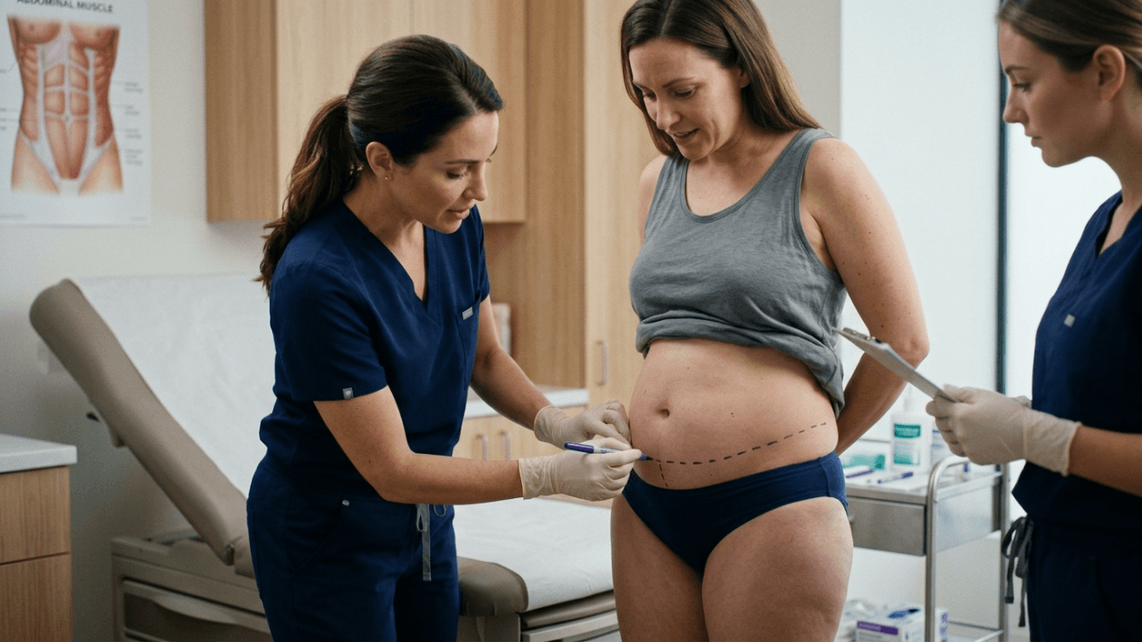
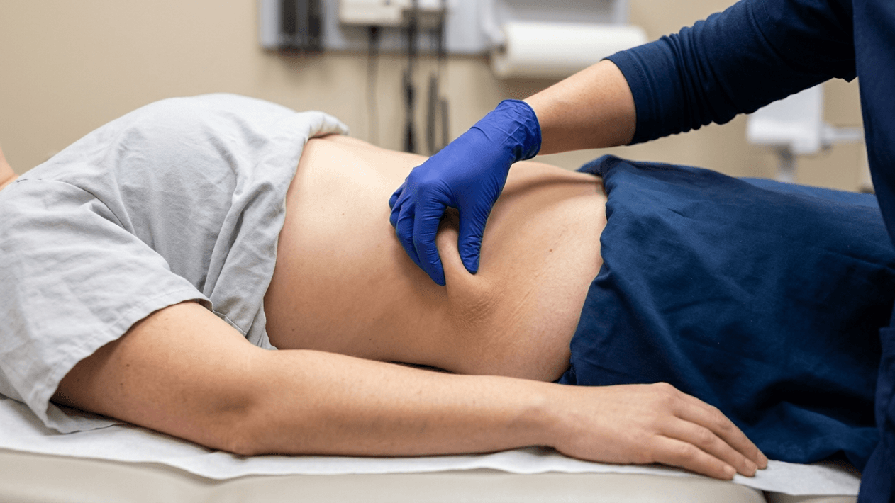
Your surgeon measures parenchymal thickness at the upper, medial, lower, and lateral poles. They evaluate skin elasticity by pinch test and stretch-mark assessment. They classify your ptosis grade using the Regnault system. They calculate your glandular-to-skin-envelope ratio. This assessment takes 30 to 45 minutes and forms the factual foundation of your entire mommy makeover plan.
Step 3: Classify Your Involution Pattern
Using the three patterns described above, your surgeon identifies whether you are a Global Deflator, Upper-Pole Collapser, or Asymmetric Involver. Each pattern maps to a distinct surgical pathway: Pattern 1 typically leads to mastopexy-only, Pattern 2 almost always requires augmentation-mastopexy, and Pattern 3 demands a hybrid approach.
Step 4: Review Before-and-After Cases Matching Your Pattern
Dr. Atalay’s Antalya clinic maintains an extensive before-and-after gallery organized by involution pattern, ptosis grade, and procedure type. Patients who review cases matching their own anatomy report clearer expectations and higher satisfaction. For instance, a 36-year-old mother of two with upper-pole collapse and Grade II ptosis can review mastopexy-only results from a similar patient — and immediately notice the difference in upper-pole contour compared with the augmentation-mastopexy results from another patient with the same ptosis grade but different glandular biology.
Step 5: Discuss Implant Selection With Precision
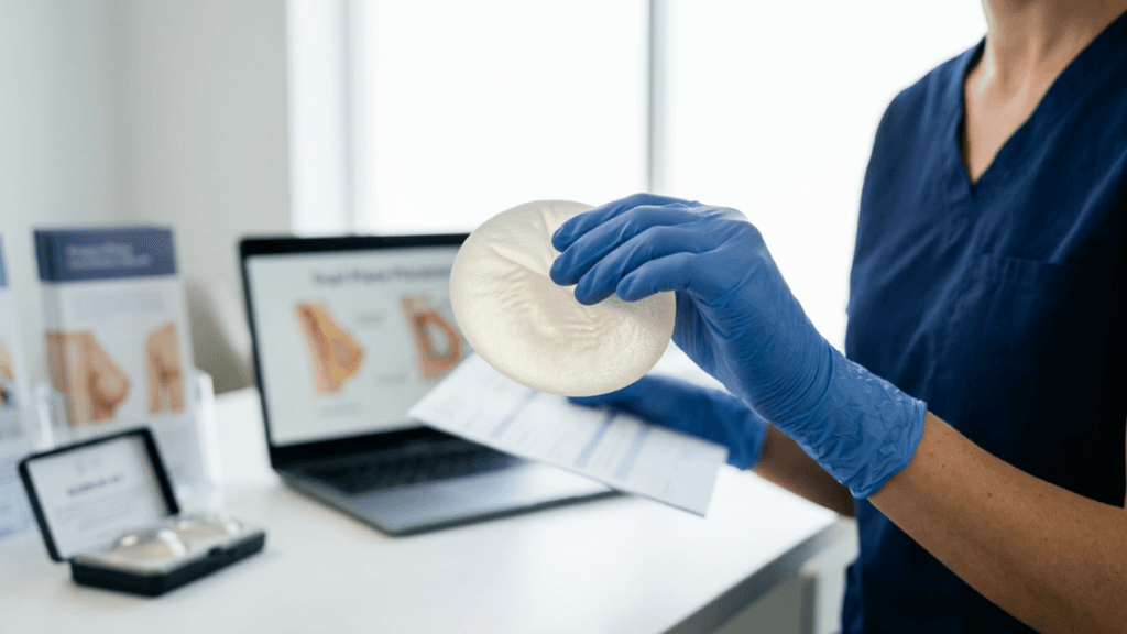
If augmentation-mastopexy is indicated, implant selection follows strict anatomical logic. Chest wall width determines base diameter. The degree of upper-pole hollowing determines projection. Dr. Atalay’s 29 years of experience with FDA- and CE-approved implants leads her toward moderate-profile devices that fill without overfilling. She deliberately avoids over-augmentation, a mistake she encounters frequently in revision patients who chose implant size based on a bra number rather than tissue capacity.
Step 6: Understand the Recovery Difference
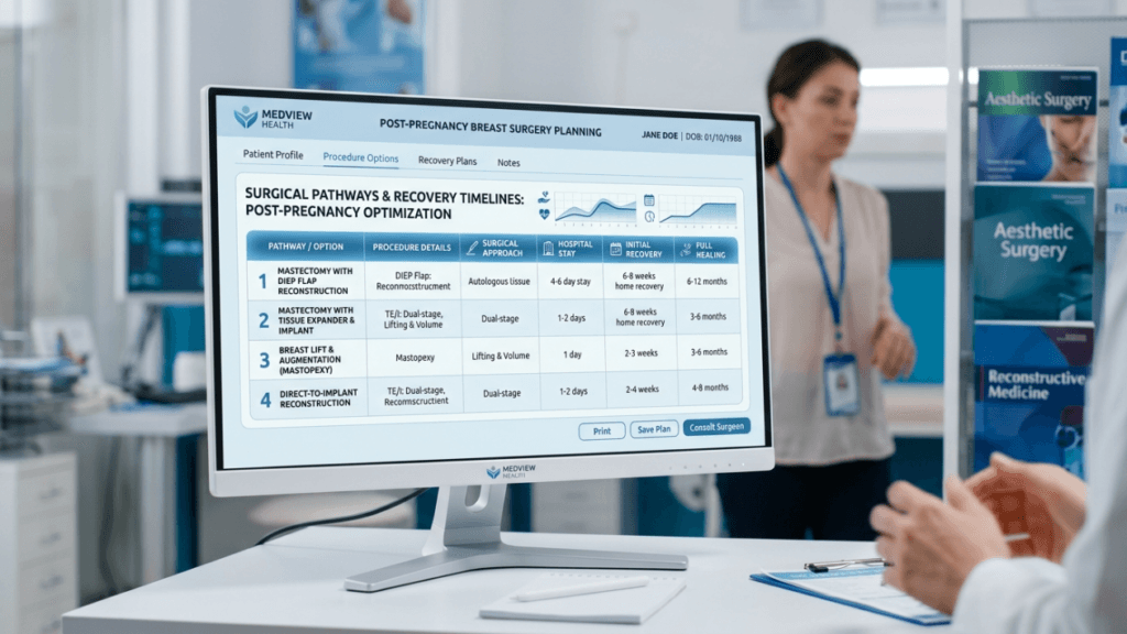
Augmentation-mastopexy recovery is longer and more involved than mastopexy alone. The dual surgical insult — implant pocket creation plus skin excision and glandular reshaping — demands four to six weeks of restricted upper-body activity. Mastopexy-only patients typically return to light activity at three weeks. Both procedures require a surgical support bra worn continuously for the first four weeks. Drain management, incision care, and swelling timelines differ substantially between the two approaches.
Patients traveling from abroad through international health tourism should plan for a minimum stay of ten to fourteen days in Antalya for augmentation-mastopexy and seven to ten days for mastopexy-only. The coastal Mediterranean climate and availability of VIP transfer and accommodation coordination make Antalya an ideal recovery destination.

The Over-Augmentation Trap: Why Bigger Is Never Better After Pregnancy
One of the most dangerous complications in post-pregnancy breast lift + implant vs. lift-only decisions is over-augmentation. Post-partum skin has already endured one cycle of extreme stretching. Asking it to carry an implant that exceeds the tissue’s structural capacity courts recurrent ptosis, widened scars, implant displacement, and bottoming-out.
Dr. Atalay addresses this proactively. During consultation, she measures your soft-tissue pinch thickness at the lower pole. If it falls below one centimeter, the implant risks being visible and palpable through the skin. She then selects the maximum implant volume that your tissue envelope can safely support — a ceiling that differs for every patient. This approach prioritizes tissue preservation and long-term structural integrity over short-term cup-size ambitions.
Clinical literature reinforces her caution. As noted in studies published in the Journal of Plastic, Reconstructive & Aesthetic Surgery, augmentation-mastopexy carries a revision rate roughly 20 times higher than primary augmentation alone. The combined forces of implant weight, tightened skin, and post-pregnancy tissue fragility create a biomechanical environment that demands conservative implant sizing.
When Mastopexy-Only Delivers Superior Results
Mastopexy-only is not a compromise. For patients with a high glandular-to-skin envelope ratio, it produces the most natural, longest-lasting result achievable. The absence of an implant eliminates the risks of capsular contracture, implant rupture, and implant-related revision surgery. The breast moves, feels, and ages like a natural breast because it is a natural breast — simply repositioned and reshaped.
Dr. Atalay’s mastopexy-only technique preserves maximal parenchymal tissue by creating an internal pedicle that lifts the glandular mound from within. This internal mastopexy technique — sometimes called a “parenchymal shaping” or “auto-augmentation” — uses the patient’s own lower-pole tissue folded upward to simulate upper-pole fullness. In select Global Deflator patients, this approach eliminates the need for an implant entirely while delivering a fuller, rounder contour than a skin-only lift.
Results from Dr. Atalay’s Antalya clinic demonstrate that patients who qualify for mastopexy-only experience fewer complications, shorter recoveries, and greater long-term satisfaction compared with augmentation-mastopexy counterparts. The key is accurate pre-operative glandular assessment that confirms adequate parenchymal volume before committing to the implant-free approach.
The Augmentation-Mastopexy Pathway: What the Combined Procedure Actually Demands
For the Upper-Pole Collapser, augmentation-mastopexy is not optional — it is anatomically necessary. The procedure combines two opposing forces: removing skin while adding volume. This tension makes it one of the most technically demanding operations in aesthetic breast surgery. Selecting a surgeon with deep experience in this specific procedure is non-negotiable.
Dr. Atalay performs augmentation-mastopexy in a single stage for the majority of post-pregnancy patients, following strict criteria: adequate soft-tissue thickness, moderate skin elasticity, and a glandular-to-envelope ratio that falls within a definable range. In patients with extremely poor skin quality — typically after multiple pregnancies and massive weight fluctuations — she may recommend a staged approach: mastopexy first, followed by implant placement three to six months later. Staging reduces tension on incisions and lowers the risk of wound-healing complications.
Implant placement for post-pregnancy augmentation-mastopexy typically uses the dual-plane technique. The upper two-thirds of the implant sits beneath the pectoralis major muscle, providing tissue coverage over the upper pole. The lower third projects behind the glandular tissue, giving the implant a natural teardrop slope. This position also reduces the risk of visible rippling through thin post-partum skin.
Real Cases From Dr. Atalay’s Antalya Clinic: Both Paths, Real Outcomes
Case A: Mastopexy-Only After Two Pregnancies
A 32-year-old patient from the United Kingdom presented after two pregnancies. Her pre-pregnancy bra size was a 34D. Post-weaning, she retained adequate glandular volume across all quadrants but developed Grade II ptosis with significant skin laxity. Her glandular-to-skin-envelope ratio measured high on assessment. Dr. Atalay performed a bilateral vertical mastopexy without implants. The parenchymal pedicle was reshaped internally to restore upper-pole contour. At six months post-operative, the patient’s breasts sit at the appropriate level on the chest wall, the upper pole shows natural fullness without any foreign material, and incision lines are fading as expected. She returned to full exercise at five weeks. The patient’s own words: “I feel like myself again, just the version from before children.”
Case B: Augmentation-Mastopexy After Three Pregnancies
A 41-year-old patient from Germany presented after three pregnancies and breastfeeding all three children. Pre-pregnancy cup size was 34B. Post-weaning involution left severe upper-pole collapse and Grade III ptosis. Her glandular assessment revealed a low parenchymal-to-envelope ratio. The skin was thin and marked with extensive striae. Dr. Atalay performed a single-stage augmentation-mastopexy using 275-cc moderate-profile silicone implants in the dual-plane position with a Wise-pattern (anchor) mastopexy. At six months, the patient achieved a full, natural-appearing breast with restored upper-pole volume, a well-positioned nipple above the inframammary fold, and symmetrical contour. Recovery extended to six weeks before full activity clearance. The patient described the outcome as transformative.
The Myth of Implants Replacing a Lift
The most harmful misconception in post-pregnancy breast surgery is the belief that large implants can compensate for sagging. This myth causes preventable revision surgeries every year. Implants add weight. Weight pulls on stretched skin. Stretched post-partum skin yields to gravity faster than never-pregnanted skin. Placing a large implant behind a sagging breast without a lift accelerates tissue descent, distorts the breast footprint, and often requires a more complex revision than the primary procedure would have been.
Conversely, the reverse myth — that a lift alone can create volume where none exists — sets equally unrealistic expectations. Mastopexy reshapes and repositions. It does not manufacture glandular tissue. If your involution pattern left you with a B-cup amount of parenchyma and a D-cup amount of skin, no lift technique can create the upper-pole convexity of a C cup. The implant becomes anatomically indispensable.
The breast lift + implant vs. lift-only decision exists precisely at the intersection of these two myths. Your glandular volume assessment cuts through both assumptions with clinical data, giving you a surgical plan anchored in your actual anatomy rather than wishful thinking.
Timing Your Surgery: Why Six Months After Weaning Is Non-Negotiable
Post-operative satisfaction in the breast lift + implant vs. lift-only pathway correlates directly with the accuracy of pre-operative assessment — and that accuracy depends on tissue stabilization. Operating before the involution process completes introduces two critical errors: the glandular volume measurement overestimates residual tissue because some incompletely involuted lobules still occupy space, and the skin envelope has not reached its final degree of laxity, meaning your surgeon tightens to a target that will shift post-operatively.
Aesthetx’s medically reviewed clinical guidance confirms the six-month stabilization window. During your consultation at Dr. Atalay’s clinic, this timeline gets discussed openly. Patients who push for earlier surgery receive a clear explanation of why waiting protects their investment. If you are still breastfeeding, your surgical plan begins after weaning — not before.
Your Mommy Makeover Action Plan: Next Steps
- Confirm timing: Ensure you are at least six months post-weaning with three months of stable body weight before scheduling a consultation.
- Schedule your glandular volume assessment: Book a consultation with a board-certified plastic surgeon who evaluates parenchymal reserve before recommending a procedure.
- Identify your involution pattern: Using the Global Deflator, Upper-Pole Collapser, and Asymmetric Involver categories, categorize your own post-pregnancy breast changes.
- Request before-and-after cases matching your pattern: Review results from patients with similar glandular-to-envelope ratios and ptosis grades.
- Discuss implant sizing limits: If augmentation-mastopexy is recommended, ask your surgeon for the maximum volume your tissue envelope can safely support long-term.
- Plan your recovery logistics: For international patients, arrange travel, accommodation, and aftercare support well in advance of your surgical date.
- Commit to your decision with clinical confidence: Your glandular assessment provides the evidence. Let anatomy — not assumption — guide your mommy makeover breast component decision.
Ready to determine whether breast lift + implant vs. lift-only is right for your post-pregnancy body? Schedule your personalized consultation with Dr. Sibel Atalay at our appointment page and take the first step toward a mommy makeover grounded in clinical precision, not guesswork.
Frequently Asked Questions About Breast Lift + Implant vs. Lift-Only After Pregnancy
How do I know if I need a breast lift + implant vs. lift-only after pregnancy?
The decision depends on your glandular-to-skin-envelope ratio. If your remaining breast tissue can adequately fill a tightened skin envelope, lift-only works. If significant upper-pole volume loss exists, augmentation-mastopexy is typically required. A clinical glandular assessment determines this.
Why does pregnancy cause breast sagging even without breastfeeding?
Pregnancy-related hormonal surges enlarge the ductal system and stretch skin and Cooper’s ligaments. After delivery, involution causes glandular tissue regression regardless of whether you breastfeed. Research confirms breastfeeding is not an independent risk factor for ptosis — pregnancy itself drives the change.
How long after weaning should I wait before breast surgery?
Breast tissue requires approximately six months after weaning to stabilize into its final shape. Operating before this window risks inaccurate glandular assessment and post-operative contour changes. Maintain a stable body weight for at least three months before your consultation.
Can implants alone fix sagging breasts after pregnancy?
No. Implants add volume but cannot tighten stretched skin. Placing implants behind a lax skin envelope without a lift typically worsens sagging over time and risks double-bubble deformity. A lift is necessary when ptosis is present regardless of whether implants are used.
Is augmentation-mastopexy riskier than mastopexy alone?
Yes. Augmentation-mastopexy combines two opposing forces — removing skin while adding volume — which increases technical complexity. Clinical studies report revision rates of 8–17% within three years compared with 5–8% for mastopexy-only. Choosing an experienced surgeon minimizes these risks.
What is the glandular-to-skin-envelope ratio?
It is a clinical measurement comparing your remaining functional breast tissue volume to the skin surface area holding it. A high ratio means sufficient glandular tissue fills the envelope. A low ratio indicates insufficient tissue, signaling the likely need for an implant alongside the lift.
Will a breast lift alone restore my pre-pregnancy upper-pole fullness?
Only if you retain adequate glandular volume in the upper pole. For women with upper-pole collapse — the most common involution pattern — a lift alone cannot create fullness where tissue no longer exists. An internal parenchymal pedicle may help, but severe hollowing requires an implant.
Why does Dr. Atalay recommend smaller implants for post-pregnancy patients?
Post-pregnancy skin has already endured significant stretching. Oversized implants increase the risk of recurrent ptosis, widened scars, and implant displacement. Dr. Atalay selects implant volumes based on your tissue capacity, prioritizing long-term structural integrity over short-term cup size.